Spider veins
Fine red, purple, or pink superficial vessels visible on the skin. These are often treated with sclerotherapy or microsclerotherapy.
Frequently asked questions for international patients with spider veins, visible surface veins, varicose veins, or suspected venous insufficiency.
Small visible veins on the legs, often called spider veins, thread veins, or surface veins, are commonly treated with sclerotherapy or microsclerotherapy. However, some patients also have larger varicose veins or underlying venous reflux, which may require a different treatment plan.
This guide explains the difference between cosmetic spider veins and medically significant varicose veins, and helps international patients plan their visit more comfortably.
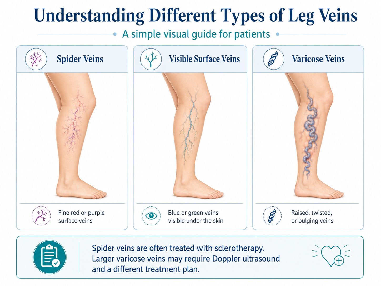
Visible leg veins are not all the same. Some are mainly cosmetic, while others may be associated with deeper venous insufficiency.
Fine red, purple, or pink superficial vessels visible on the skin. These are often treated with sclerotherapy or microsclerotherapy.
More noticeable blue or green veins under the skin. Treatment depends on their size and whether reflux is present.
Larger bulging, raised, twisted, or rope-like veins. These usually require Doppler ultrasound before deciding on treatment.
Sclerotherapy is a minimally invasive treatment used for visible small veins and spider veins on the legs. A special medication is injected into the targeted veins with fine needles. Over time, the treated veins close and gradually become less visible.
Microsclerotherapy is a refined form of sclerotherapy used for very small superficial veins. It is commonly used for cosmetic spider veins and thread veins on the legs.
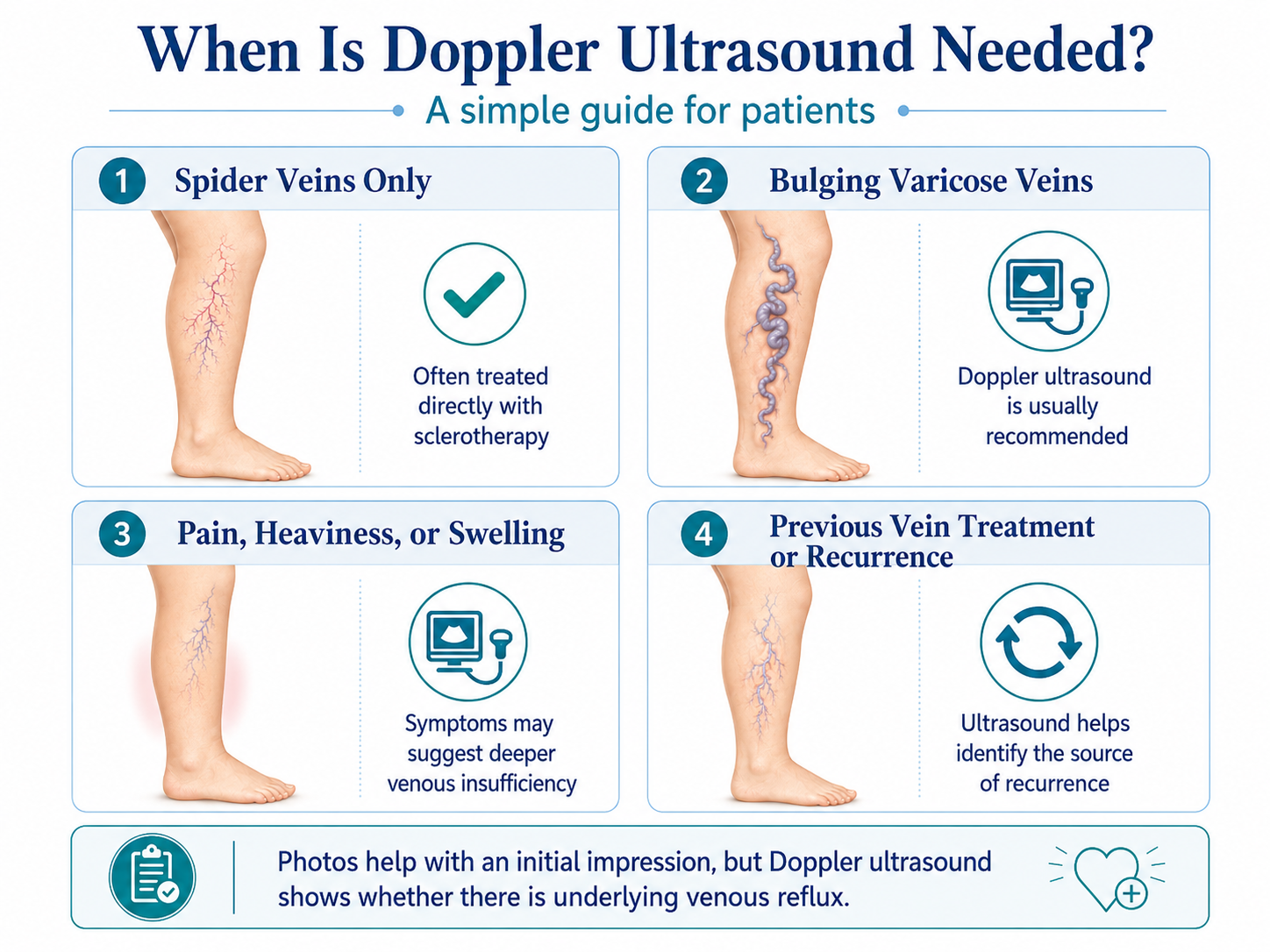
Not always. Small spider veins and superficial visible veins are often suitable for sclerotherapy. However, if there are larger bulging varicose veins, leg swelling, heaviness, pain, skin discoloration, or visible rope-like veins, a venous Doppler ultrasound may be needed first.
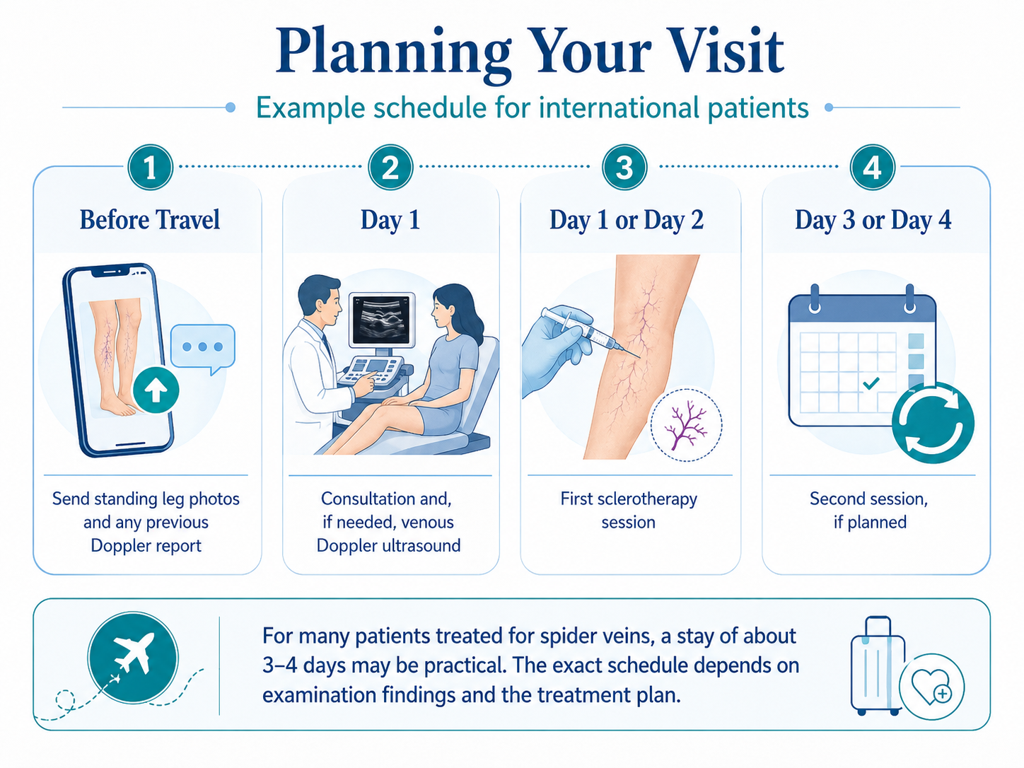
Yes. Clear photos taken while standing are very helpful. Photos of both legs from the front, back, and sides can help provide an initial impression of the visible veins and the likely treatment direction.
The number of sessions depends on the extent and density of the veins. For many patients with spider veins, 2 to 3 sessions may be enough for an initial treatment plan. In mild cases, fewer sessions may be sufficient, while more extensive cases may require additional sessions.
No. Treatment is not usually calculated strictly by separate areas such as front leg, back leg, or one small region. During one session, both legs can be treated, depending on the extent of visible veins and the safe amount of sclerosant that can be used in one session.
A venous Doppler ultrasound is important because visible veins on the surface may sometimes be only the outer sign of an underlying reflux problem.
Doppler ultrasound is commonly recommended in the following situations:
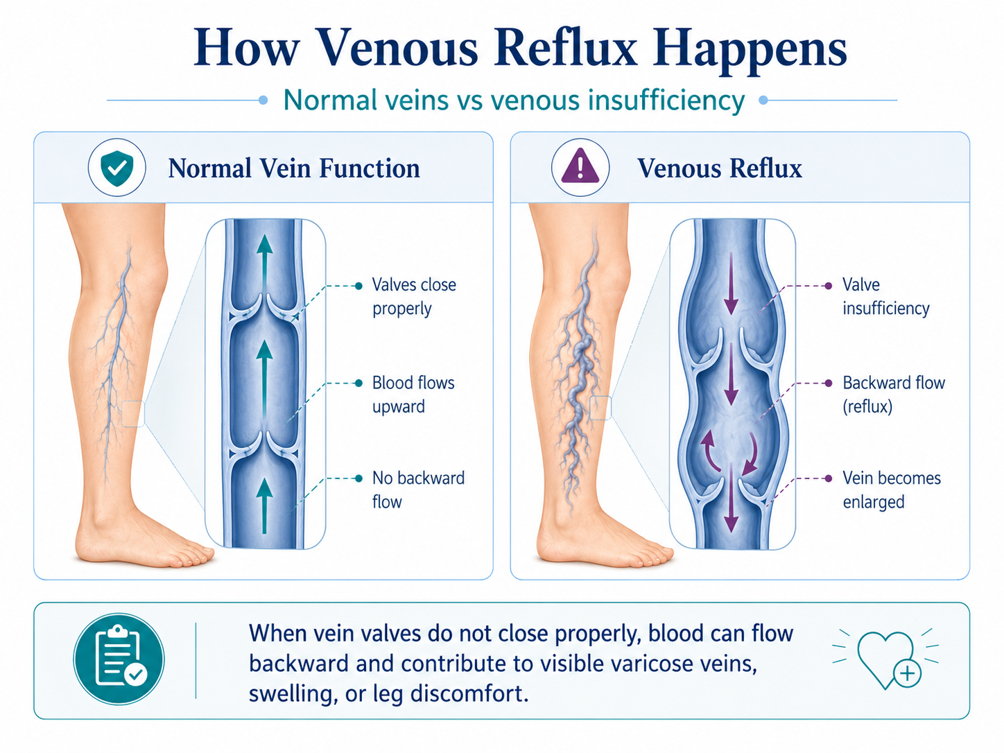
Venous reflux means that the vein valves do not close properly and blood flows backward instead of upward. Over time, this may contribute to visible varicose veins, swelling, leg discomfort, or recurrence after previous treatment.
If reflux is present in veins such as the great saphenous vein, small saphenous vein, accessory saphenous veins, or perforator veins, treating only the visible surface veins may not be enough.
If you have larger, raised, bulging, or rope-like varicose veins, the treatment may be different from simple spider vein sclerotherapy.
These veins are often associated with an underlying reflux problem. In such cases, a venous Doppler ultrasound examination is usually necessary to understand the source of the problem and choose the correct treatment plan.
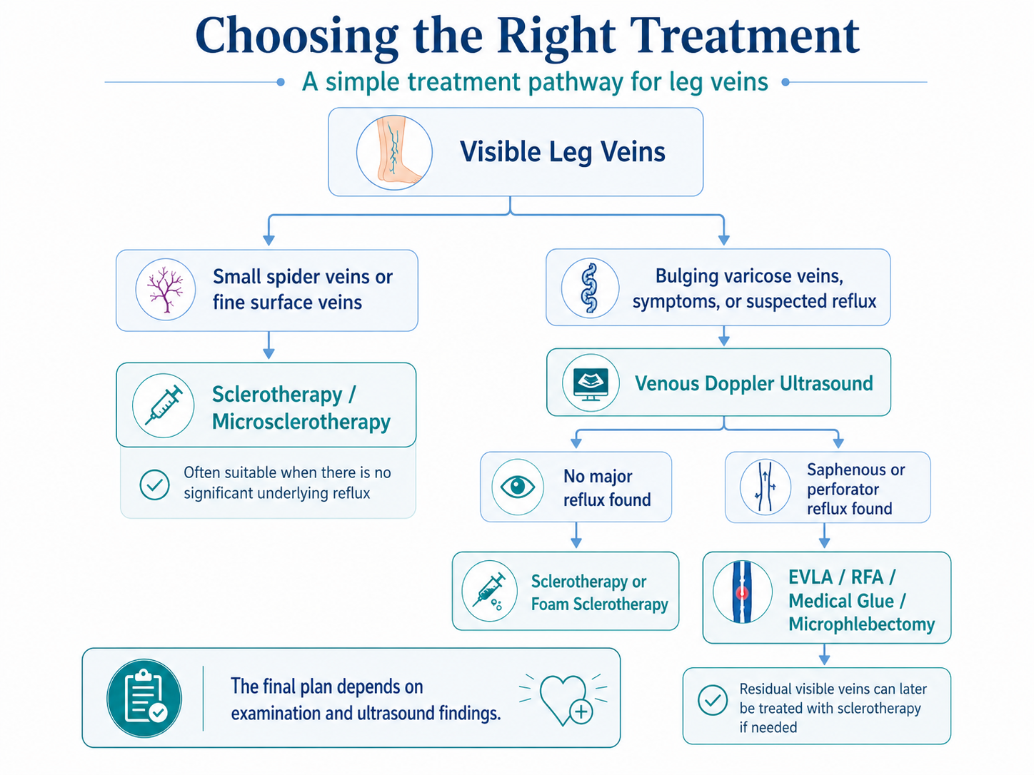
If there is significant reflux in the saphenous veins, sclerotherapy alone may not be sufficient. In such cases, the refluxing vein usually needs to be treated first. After the main reflux source is treated, any remaining smaller visible veins can be treated with sclerotherapy if needed.
Depending on the Doppler ultrasound findings, treatment options may include:
The final treatment plan depends on the ultrasound findings and in-person examination.
Sometimes yes, but not always.
If the patient has only small spider veins or fine superficial veins, treatment may often start directly with sclerotherapy after examination.
However, if there are larger varicose veins or signs of venous insufficiency, Doppler ultrasound should usually be performed first. If a deeper reflux problem is identified, the priority is usually to treat the main refluxing vein. Spider veins and smaller remaining veins can then be treated in the same treatment period or in later sessions.
If two sessions are planned, a stay of about 3 to 4 days is often practical. Sessions can usually be arranged with 1 to 2 days between them.
If a third session is needed, it may sometimes be possible to perform it 1 to 2 days after the second session, depending on the patient’s condition and schedule.
If Doppler ultrasound shows significant reflux or larger varicose veins, the treatment plan may be more comprehensive. In that case, the required stay may be longer depending on the type of treatment and whether additional sessions are planned.
For this reason, international patients are advised to send clear standing photos of both legs and, if available, any previous venous Doppler ultrasound report before travelling.
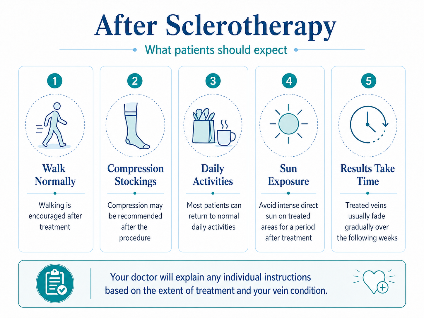
Yes. Walking is encouraged after treatment. Most patients can return to daily activities quickly.
Most patients can travel after treatment, but the timing depends on the extent of the procedure and the travel distance. It is generally better not to plan an excessively rushed schedule.
Compression stockings are commonly recommended after sclerotherapy. The duration of use depends on the extent of treatment and the patient’s vein condition.
Sclerotherapy can be performed in different seasons, but many patients prefer cooler months because compression stockings are more comfortable and sun exposure is easier to avoid.
It is better to avoid direct intense sun exposure on treated areas for a period after treatment, as this may help reduce the risk of pigmentation.
Treated veins do not disappear immediately. They usually fade gradually over the following weeks. Some veins may temporarily look darker before improving.
Sclerotherapy can significantly reduce visible spider veins, but it is not realistic to promise complete disappearance of every small vessel. Some veins may require more than one session, and new veins can appear over time.
Photos are very helpful for an initial impression, especially for visible spider veins and superficial varicose veins. However, photos cannot show the function of the deeper veins or whether reflux is present.
For this reason, photos can help estimate the possible treatment direction, but the final plan can only be made after medical examination and, when necessary, venous Doppler ultrasound.
Patients who wish to receive a preliminary opinion may send:
After reviewing the available information, it may be possible to provide a preliminary idea about the likely treatment direction and the estimated number of sessions. A final decision is made after in-person examination.
This page is intended for general patient information. The final treatment plan always depends on medical examination and, when necessary, venous Doppler ultrasound findings.
For questions about planning your visit, contact the center.